Mice whose mitochondria were briefly stressed before birth grew up with hearts resistant to a chemotherapy drug that causes heart failure. A La Jolla study pinpoints the molecular chain behind the protection.
The Salk Institute study, published Sept. 4 in the journal Science Advances, explores a process called mitohormesis. The concept is counterintuitive: a small dose of stress inside mitochondria during embryonic development can reprogram cells to be more resilient for life.
Gerald Shadel, professor and holder of the Audrey Geisel Chair in Biomedical Science at the Salk Institute, led the research. Matthew Donnelly, an MD/PhD graduate student in Shadel's lab, is the study's first author.
The team temporarily lowered levels of SOD2, an antioxidant enzyme that removes a reactive molecule called superoxide from mitochondria, during mouse embryonic development. They restored the enzyme before birth. By six weeks old, those mice showed changes in 201 genes tied to energy production, antioxidant defense and fat metabolism, according to the study.
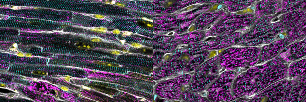
When the researchers later treated the mice with doxorubicin, a chemotherapy drug known to damage mitochondria and cause heart failure, the stressed-in-utero mice were protected from the drug's cardiac toxicity. A control group was not.
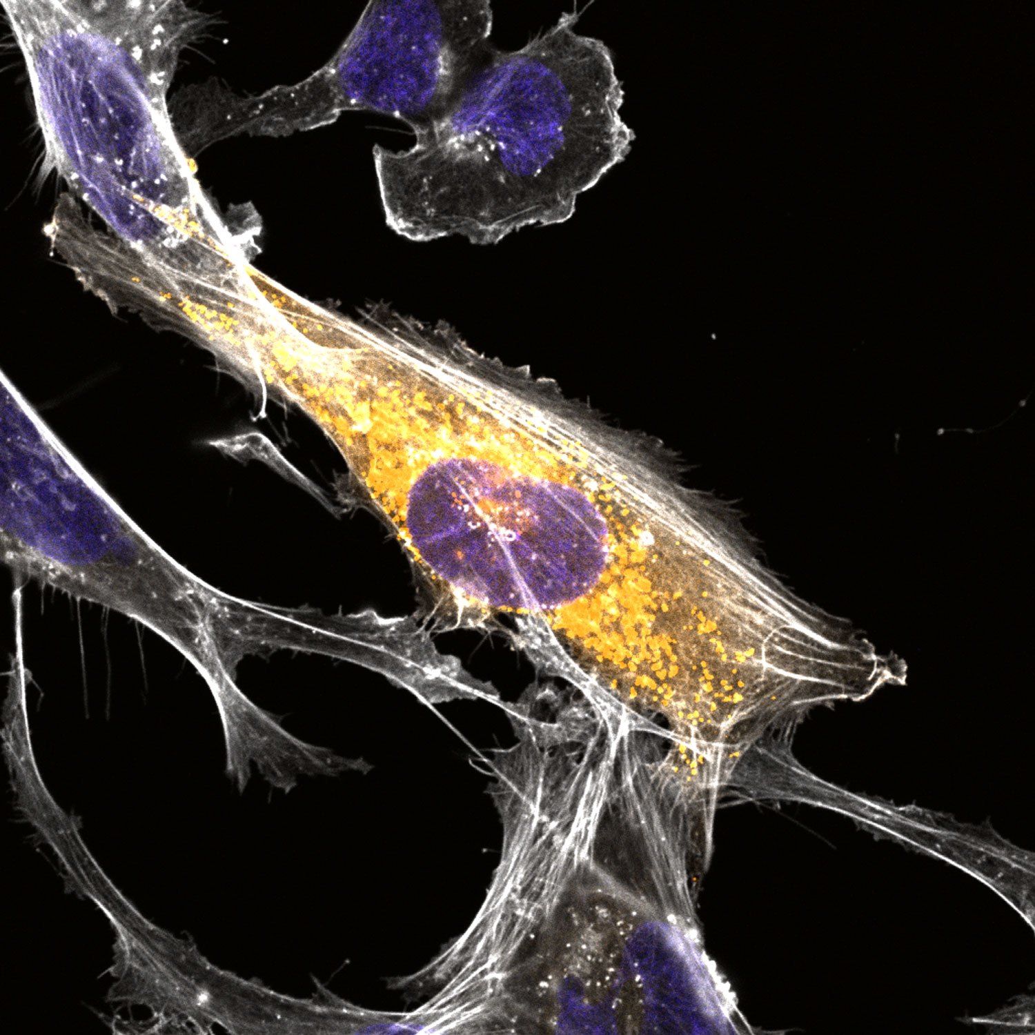
The team then moved to mouse embryonic fibroblasts to trace the mechanism. They found that superoxide buildup blocked a key enzyme in the energy-production cycle, causing citrate to accumulate inside mitochondria. That citrate leaked out and was converted into acetyl-CoA, a molecule that triggers epigenetic changes. Those changes made the cells durably more resistant to future stress.
Citrate, the researchers concluded, acts as a "second messenger" for superoxide, carrying the stress signal from inside the mitochondria to the cell's gene-regulation machinery.
"Antioxidant therapies have been developed to mitigate damaging effects of ROS, but they have largely failed in clinical trials … A better therapeutic approach to just targeting one reactive oxygen species at a time might be to enact a more nuanced global program like mitohormesis that affects both mitochondria and several antioxidant systems simultaneously," Shadel said in the Salk Institute's Sept. 4 press release.
The finding builds on Shadel's earlier work. In 2018, his lab demonstrated mitohormesis in mammals using the same mouse strain. That study focused on the liver and found embryonic mitochondrial stress led mice to grow more mitochondria. The new study extends the same approach to the heart. Shadel received the 2026 Charles L. Hoppel Prize from Case Western Reserve University in May for his contributions to mitochondrial biology.
The study does not suggest people should try to create oxidative stress or take citrate supplements. The strongest evidence came from mice and mouse cells. Co-authors include researchers from the Salk Institute and Asa B. Gustafsson of UC San Diego. The National Institutes of Health (NIH), the Arnold and Mabel Beckman Foundation, the Cancer Research Institute and the Chan Zuckerberg Initiative funded the work.
The Salk team's next steps include testing whether mitohormesis can be triggered after embryonic development and whether the findings hold in human tissue models.